Beranda
/ Foot Muscles Mri : Magnetic Resonance Imaging Mri Image Showing Foot Muscles And Download Scientific Diagram - 31 the plantar intrinsic foot muscles consist of four layers of muscles deep to the plantar aponeurosis.
Foot Muscles Mri : Magnetic Resonance Imaging Mri Image Showing Foot Muscles And Download Scientific Diagram - 31 the plantar intrinsic foot muscles consist of four layers of muscles deep to the plantar aponeurosis.
Insurance Gas/Electricity Loans Mortgage Attorney Lawyer Donate Conference Call Degree Credit Treatment Software Classes Recovery Trading Rehab Hosting Transfer Cord Blood Claim compensation mesothelioma mesothelioma attorney Houston car accident lawyer moreno valley can you sue a doctor for wrong diagnosis doctorate in security top online doctoral programs in business educational leadership doctoral programs online car accident doctor atlanta car accident doctor atlanta accident attorney rancho Cucamonga truck accident attorney san Antonio ONLINE BUSINESS DEGREE PROGRAMS ACCREDITED online accredited psychology degree masters degree in human resources online public administration masters degree online bitcoin merchant account bitcoin merchant services compare car insurance auto insurance troy mi seo explanation digital marketing degree floridaseo company fitness showrooms stamfordct how to work more efficiently seowordpress tips meaning of seo what is an seo what does an seo do what seo stands for best seotips google seo advice seo steps, The secure cloud-based platform for smart service delivery. Safelink is used by legal, professional and financial services to protect sensitive information, accelerate business processes and increase productivity. Use Safelink to collaborate securely with clients, colleagues and external parties. Safelink has a menu of workspace types with advanced features for dispute resolution, running deals and customised client portal creation. All data is encrypted (at rest and in transit and you retain your own encryption keys. Our titan security framework ensures your data is secure and you even have the option to choose your own data location from Channel Islands, London (UK), Dublin (EU), Australia.
Foot Muscles Mri : Magnetic Resonance Imaging Mri Image Showing Foot Muscles And Download Scientific Diagram - 31 the plantar intrinsic foot muscles consist of four layers of muscles deep to the plantar aponeurosis.. Mri with hardware in foot? Epidemiology of tuberculosis etiology tuberculous spondylodiscitis clinical manifestations review of imaging findings: This is a 30 year old with swelling on the lateral aspect of foot with evidence of soft tissue lesion in relation to the lateral aspect of the talus which appears isointense to the muscles on t1 and t2 weighted images & appears elongated extending from the anterosuperior calcaneum to the base of. There can't be any metal in the room, not just where you have the mri. Posted by radiologyer at 8:12 am.
It must be placed in the center of the magnet, to obtain homogeneous fat. They act collectively to stabilise the arches of the foot, and individually to control movement of the digits. The muscles of the dorsum of the foot are a group of two muscles, which together represent the dorsal foot musculature. As a result, during walking the body's center of gravity normally fluctuates only 5cm in both vertical and lateral directions. However, on mri images, no muscular abnormalities were detected.
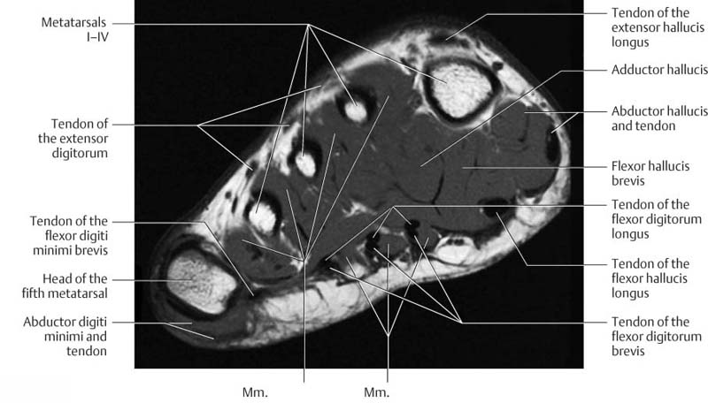
Mri Lower Extremities Leg Cedars Sinai from www.cedars-sinai.org The abductor digiti minimi muscle is on the lateral side of the foot and contributes to the large lateral plantar eminence on the sole. Synovitis, tenosynovitis, bursitis, and ganglion cysts) > congenital and developmental conditions( eg.dysplasia, tarsal coalition). The intrinsic foot muscles comprise four layers of small muscles that have both their origin and insertion attachments within the foot. Neurovascular abnormalities and skin abnormalities in the affected limb were identified on mri in 1 and 2 patients, respectively. 12 photos of the foot muscle anatomy mri. The muscle that removes the big toe (m.abductor hallucis) lies superficially along the medial edge of the foot. They act collectively to stabilise the arches of the foot, and individually to control movement of the digits. It begins with short tendon bundles on the medial surface of the calcaneus calcaneus, fleshy bundles on the lower retentive flexor.
This means that the little toe can only be extended by the extensor digitorum longus muscle only.
The purpose of this study was to investigate the relationship of muscle mri findings and gait disturbance in myotonic dystrophy type 1 (dm1) patients. 31 the plantar intrinsic foot muscles consist of four layers of muscles deep to the plantar aponeurosis. Synovitis, tenosynovitis, bursitis, and ganglion cysts) > congenital and developmental conditions( eg.dysplasia, tarsal coalition). Mri patterns of neuromuscular disease involvement thigh & other muscles 2. Where you get the potential for problems with. Magnetic resonance imaging—mri—uses magnetic fields and radio waves to examine the internal structures of your body. ► shoulder ► elbow ► wrist ► finger ► thumb. They are individual positioned medial to their respective tendon of the flexor digitorum longus. However, on mri images, no muscular abnormalities were detected. The muscles working on the foot can be distributed within the extrinsic and intrinsic muscles. The muscles of the dorsum of the foot are a group of two muscles, which together represent the dorsal foot musculature. Indications for foot mri scan. Posted by radiologyer at 8:12 am.
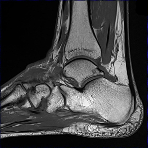
The intrinsic foot muscles (ifm) are the main local stabilizers of the foot and are part of the active and neural subsystems that constitute the foot core. In addition, an image of all the muscles of the back and plantar part of the foot, all tendons and tendon ligaments, blood vessels and nerves are obtained. Indications for foot mri scan. 12 photos of the foot muscle anatomy mri. Top suggestions for foot muscle anatomy mri.
Mri Of The Foot Radiology Key from i0.wp.com This is a 30 year old with swelling on the lateral aspect of foot with evidence of soft tissue lesion in relation to the lateral aspect of the talus which appears isointense to the muscles on t1 and t2 weighted images & appears elongated extending from the anterosuperior calcaneum to the base of. Musculoskeletal system | muscle structure and function. Mri with hardware in foot? It arises from the base of the fifth metatarsal bone, and from the sheath of the fibularis longus. Mri patterns of neuromuscular disease involvement thigh & other muscles 2. The muscles of the dorsum of the foot are a group of two muscles, which together represent the dorsal foot musculature. There can't be any metal in the room, not just where you have the mri. Where you get the potential for problems with.
It begins with short tendon bundles on the medial surface of the calcaneus calcaneus, fleshy bundles on the lower retentive flexor.
It begins with short tendon bundles on the medial surface of the calcaneus calcaneus, fleshy bundles on the lower retentive flexor. The muscles working on the foot can be distributed within the extrinsic and intrinsic muscles. Muscles of the foot are located on its rear and on the sole. Epidemiology of tuberculosis etiology tuberculous spondylodiscitis clinical manifestations review of imaging findings: Lateral and medial processes of calcaneal tuberosity, and band of connective tissue connecti. This means that the little toe can only be extended by the extensor digitorum longus muscle only. As a result, during walking the body's center of gravity normally fluctuates only 5cm in both vertical and lateral directions. Where you get the potential for problems with. Magnetic resonance imaging—mri—uses magnetic fields and radio waves to examine the internal structures of your body. Synovitis, tenosynovitis, bursitis, and ganglion cysts) > congenital and developmental conditions( eg.dysplasia, tarsal coalition). Musculoskeletal system | muscle structure and function. In addition, an image of all the muscles of the back and plantar part of the foot, all tendons and tendon ligaments, blood vessels and nerves are obtained. The purpose of this study was to investigate the relationship of muscle mri findings and gait disturbance in myotonic dystrophy type 1 (dm1) patients.
The muscles acting on the foot can be divided into two distinct groups; Indications for foot mri scan. The purpose of this study was to investigate the relationship of muscle mri findings and gait disturbance in myotonic dystrophy type 1 (dm1) patients. There are 10 intrinsic muscles located in the sole of the foot. ► shoulder ► elbow ► wrist ► finger ► thumb.
Ankle And Foot Radiology Key from radiologykey.com The muscles acting on the foot can be divided into two distinct groups; Foot ulceration can subsequently lead to infections, such as cellulitis and osteomyelitis, and this may eventually the mri examination includes special attention for positioning of the foot. Muscles of the foot are located on its rear and on the sole. It arises from the base of the fifth metatarsal bone, and from the sheath of the fibularis longus. Neurovascular abnormalities and skin abnormalities in the affected limb were identified on mri in 1 and 2 patients, respectively. Involved early gray = muscle: ► shoulder ► elbow ► wrist ► finger ► thumb. Indications for foot mri scan.
In addition, an image of all the muscles of the back and plantar part of the foot, all tendons and tendon ligaments, blood vessels and nerves are obtained.
As a result, during walking the body's center of gravity normally fluctuates only 5cm in both vertical and lateral directions. The muscles of the dorsum of the foot are a group of two muscles, which together represent the dorsal foot musculature. 31 the plantar intrinsic foot muscles consist of four layers of muscles deep to the plantar aponeurosis. Posted by radiologyer at 8:12 am. Top suggestions for foot muscle anatomy mri. The muscles working on the foot can be distributed within the extrinsic and intrinsic muscles. Involved early gray = muscle: The abductor digiti minimi muscle is on the lateral side of the foot and contributes to the large lateral plantar eminence on the sole. This is a 30 year old with swelling on the lateral aspect of foot with evidence of soft tissue lesion in relation to the lateral aspect of the talus which appears isointense to the muscles on t1 and t2 weighted images & appears elongated extending from the anterosuperior calcaneum to the base of. The purpose of this study was to investigate the relationship of muscle mri findings and gait disturbance in myotonic dystrophy type 1 (dm1) patients. 12 photos of the foot muscle anatomy mri. They act collectively to stabilise the arches of the foot, and individually to control movement of the digits. Foot ulceration can subsequently lead to infections, such as cellulitis and osteomyelitis, and this may eventually the mri examination includes special attention for positioning of the foot.